Address: Main Building, Eftimie Murgu Square, Nr. 2, 300041, Timişoara
Phone: 0256/204250
E-mail: flavia.zara@umft.ro
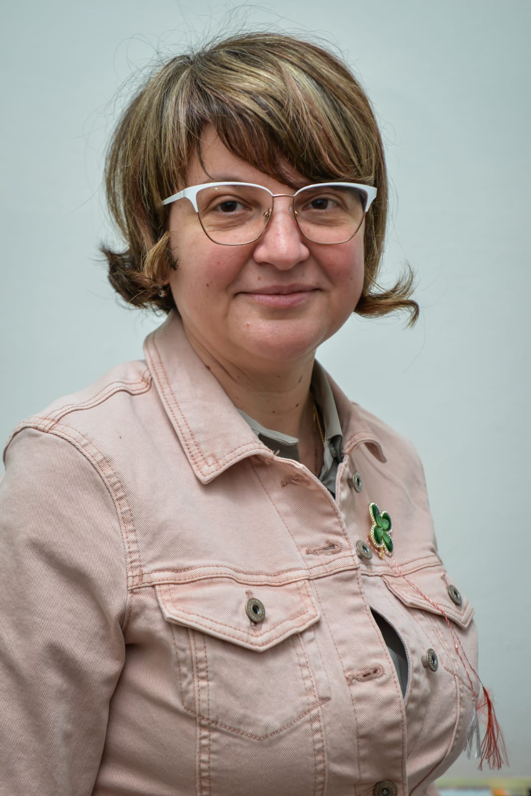
Head of the Academic Department
Members
Short history. The discipline of Histology was founded in 1945, having as initiator, organizer and coordinator Prof. Dr. Şerban Brătianu, the dean of the Faculty of Medicine at that time. The activity of the discipline included from the beginning two distinct development directions, didactic and research, the last being intensely publicized through professor Brătianu’s publications in specialized literature at that time. The modernization of the histology discipline has been continuously carried out, being evident since 1995, when the current head of discipline, Professor Marius Raica reorganized the material basis necessary for the teaching activity and initiated the organization of a research center equipped to international standards, namely the Angiogenesis Research Center, www.angiotm.com.
Activity specific. Currently, the discipline of Hisology includes three main development directions: didactics, research and mentoring / tutoring / volunteering activities.
The didactic activity has as main field the study of normal cells and tissues through classical and modern microscopic techniques, as well as the objective and transparent evaluation of the students’ aptitudes through a year examination structured in two distinct directions: theoretical and practical.
The didactic activities of the discipline are addressed to the Romanian students of the second year of Medicine, the first year of Dental Medicine, General Medicine Assistance (Timisoara, Lugoj, Deva) as well as for the students from the corresponding years from the English and French program. The first-year students benefit of the teaching activities of the discipline also from the Nutrition and Dietetics programs. Teachers and students develop their activities in an amphyteatre and four practical laboratories, two of them being equipped with microscopes and the other two being recently reorganized into two virtual microscopy laboratories. These are equipped with a system for scanning microscopic slides and storing them on electronic support followed by their computerized presentation. All these facilities are accessible to students during practical laboratories at the University as well as from other locations based on a personalized account. The lecture support includes powerpoint presentations, books written by the members of the discipline and protocols of practical laboratory realised by the teachers in collaboration with the students included in the tutoring program. Details regarding the subject sheet, the schedule, the consultation program, the bibliography, the exam results are displayed on the subject’s notice board.
The scientific research is carried out within the Research Center in Angiogenesis which includes the following compartments:
- Primary tissue processing laboratory
- Classical microscopy laboratory with fluorescence in ApoTome system and virtual microscopy
- Microscopy laboratory with microdissection laser.
- MicroArray Tissue Laboratory
- Automated immunohistochemistry laboratory
- Laboratory of molecular pathology: extraction and DNA analysis; RNA, in situ hybirdization with chromogen and fluorescence, classical RT PCR techniques and in TaqMan Array system.
- Laboratory of experimental medicine on model of chorioalantoid membrane
Details of the equipment of each laboratory can be found at the http://www.angiotm.com/labs.html
Priority areas of research include: malignant tumor pathology, angiogenesis and normal and tumor lymphangiogenesis, molecular profile of malignant tumors and its involvement in diagnosis and therapy, implementation and use of new experimental models. The tumor pathology addressed concerns malignant head and neck tumors, clear cell carcinomas, urothelial malignant tumors, breast cancer, pituitary adenomas. The secondary fields of research include the microscopic and molecular approach of placental pathology, atheroma plaque, peripheral vascular changes, microscopic lesions induced by diabetes mellitus on experimental model, posttraumatic regeneration of peripheral nerve by addition of adipose tissue, corneal ocular pathology.
The tutoring/mentoring/volunteering activity includes the selection of students with outstanding skills and performances who want to continue their activity in the histology discipline after passing the final exam. Based on the concept of “students learn students”, the tutoring/mentoring program offers the possibility to the student to become a teacher for his colleagues under the guidance of a tenured teacher of the discipline from the time of his/her student days. Moreover, the collaboration of the student tutor-teacher determines the improvement of the student-teacher communication as well as the facilitation of the teacher’s adaptation to the dynamic needs of the student.
- Didactic activity
The implementation of the study of histology in the computerized system determined the uniformity of the transmission of information to the students involved in the study of histology. The evaluation of the students in the computerized system determined the increase of the promoteability by approximately 20% at the first presentation compared to the classical evaluation system. This evaluation system has also eliminated many controversial aspects of classical examination and is characterised by transparency and objective results.
- The research and dissemination of research results can be viewed using the link http://www.angiotm.com/articles.html
Tutoring/mentoring/volunteering activity. It materialized by the selection of 4 permanent tutors, students in years III-VI, as well as an assistant professor former tutor in the discipline of histology, currently a resident physician and member of our team. Tutors are also involved in research activities. Details can be found in the http://www.angiotm.com/departament.html section “Students”. Writing an e-book by tutors from the English section, A Histology Atlas for Students made by Students, Part I-General Histology, authors Erik Jan Dijkstra; Andreas Zoric ; Andreas Salagean, a book that is currently used as an electronic support for the protocols of practical histology works for foreign students, can be viewed and read online using the link http://umftebooks.ro/ after the prior realization of a user account.

